↓ Skip to main content

The identification and neurochemical characterization of central neurons that target parasympathetic preganglionic neurons involved in the regulation of choroidal blood flow in the rat eye using…

Overview of attention for article published in Frontiers in Neuroanatomy, June 2015
Altmetric Badge

About this Attention Score

  • Above-average Attention Score compared to outputs of the same age (52nd percentile)
  • Average Attention Score compared to outputs of the same age and source

Mentioned by

twitter
3 X users

Citations

dimensions_citation
16 Dimensions

Readers on

mendeley
36 Mendeley
You are seeing a free-to-access but limited selection of the activity Altmetric has collected about this research output. Click here to find out more.
Title
The identification and neurochemical characterization of central neurons that target parasympathetic preganglionic neurons involved in the regulation of choroidal blood flow in the rat eye using pseudorabies virus, immunolabeling and conventional pathway tracing methods
Published in
Frontiers in Neuroanatomy, June 2015
DOI 10.3389/fnana.2015.00065
Pubmed ID
Authors

Chunyan Li, Malinda E. C. Fitzgerald, Nobel Del Mar, Sherry Cuthbertson-Coates, Mark S. LeDoux, Suzhen Gong, James P. Ryan, Anton Reiner

Abstract

The choroidal blood vessels of the eye provide the main vascular support to the outer retina. These blood vessels are under parasympathetic vasodilatory control via input from the pterygopalatine ganglion (PPG), which in turn receives its preganglionic input from the superior salivatory nucleus (SSN) of the hindbrain. The present study characterized the central neurons projecting to the SSN neurons innervating choroidal PPG neurons, using pathway tracing and immunolabeling. In the initial set of studies, minute injections of the Bartha strain of the retrograde transneuronal tracer pseudorabies virus (PRV) were made into choroid in rats in which the superior cervical ganglia had been excised (to prevent labeling of sympathetic circuitry). Diverse neuronal populations beyond the choroidal part of ipsilateral SSN showed transneuronal labeling, which notably included the parvocellular part of the paraventricular nucleus of the hypothalamus (PVN), the periaqueductal gray, the raphe magnus (RaM), the B3 region of the pons, A5, the nucleus of the solitary tract (NTS), the rostral ventrolateral medulla (RVLM), and the intermediate reticular nucleus of the medulla. The PRV+ neurons were located in the parts of these cell groups that are responsive to systemic blood pressure signals and involved in systemic blood pressure regulation by the sympathetic nervous system. In a second set of studies using PRV labeling, conventional pathway tracing, and immunolabeling, we found that PVN neurons projecting to SSN tended to be oxytocinergic and glutamatergic, RaM neurons projecting to SSN were serotonergic, and NTS neurons projecting to SSN were glutamatergic. Our results suggest that blood pressure and volume signals that drive sympathetic constriction of the systemic vasculature may also drive parasympathetic vasodilation of the choroidal vasculature, and may thereby contribute to choroidal baroregulation during low blood pressure.
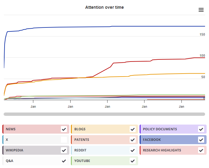
Timeline

Login to access the full chart related to this output.

If you don’t have an account, click here to discover Explorer

X Demographics

X Demographics

The data shown below were collected from the profiles of 3 X users who shared this research output. Click here to find out more about how the information was compiled.
As of 1 July 2024, you may notice a temporary increase in the numbers of X profiles with Unknown location. Click here to learn more.
Mendeley readers

Mendeley readers

The data shown below were compiled from readership statistics for 36 Mendeley readers of this research output. Click here to see the associated Mendeley record.

Geographical breakdown

Country Count As %
Unknown 36 100%

Demographic breakdown

Readers by professional status Count As %
Researcher 7 19%
Other 4 11%
Student > Ph. D. Student 4 11%
Professor 4 11%
Student > Bachelor 3 8%
Other 6 17%
Unknown 8 22%
Readers by discipline Count As %
Neuroscience 9 25%
Agricultural and Biological Sciences 5 14%
Medicine and Dentistry 5 14%
Biochemistry, Genetics and Molecular Biology 2 6%
Unspecified 2 6%
Other 3 8%
Unknown 10 28%
Attention Score in Context

Attention Score in Context

This research output has an Altmetric Attention Score of 3. This is our high-level measure of the quality and quantity of online attention that it has received. This Attention Score, as well as the ranking and number of research outputs shown below, was calculated when the research output was last mentioned on 18 June 2015.
All research outputs
#13,361,781
of 22,808,725 outputs
Outputs from Frontiers in Neuroanatomy
#563
of 1,159 outputs
Outputs of similar age
#126,078
of 267,792 outputs
Outputs of similar age from Frontiers in Neuroanatomy
#22
of 41 outputs
Altmetric has tracked 22,808,725 research outputs across all sources so far. This one is in the 41st percentile – i.e., 41% of other outputs scored the same or lower than it.
So far Altmetric has tracked 1,159 research outputs from this source. They typically receive more attention than average, with a mean Attention Score of 9.0. This one has gotten more attention than average, scoring higher than 50% of its peers.
Older research outputs will score higher simply because they've had more time to accumulate mentions. To account for age we can compare this Altmetric Attention Score to the 267,792 tracked outputs that were published within six weeks on either side of this one in any source. This one has gotten more attention than average, scoring higher than 52% of its contemporaries.
We're also able to compare this research output to 41 others from the same source and published within six weeks on either side of this one. This one is in the 46th percentile – i.e., 46% of its contemporaries scored the same or lower than it.