↓ Skip to main content

Septo-temporal distribution and lineage progression of hippocampal neurogenesis in a primate (Callithrix jacchus) in comparison to mice

Overview of attention for article published in Frontiers in Neuroanatomy, June 2015
Altmetric Badge

About this Attention Score

  • Good Attention Score compared to outputs of the same age (72nd percentile)
  • Good Attention Score compared to outputs of the same age and source (68th percentile)

Mentioned by

twitter
10 X users

Readers on

mendeley
58 Mendeley
You are seeing a free-to-access but limited selection of the activity Altmetric has collected about this research output. Click here to find out more.
Title
Septo-temporal distribution and lineage progression of hippocampal neurogenesis in a primate (Callithrix jacchus) in comparison to mice
Published in
Frontiers in Neuroanatomy, June 2015
DOI 10.3389/fnana.2015.00085
Pubmed ID
Authors

Irmgard Amrein, Michael Nosswitz, Lutz Slomianka, R. Maarten van Dijk, Stefanie Engler, Fabienne Klaus, Olivier Raineteau, Kasum Azim

Abstract

Adult born neurons in the hippocampus show species-specific differences in their numbers, the pace of their maturation and their spatial distribution. Here, we present quantitative data on adult hippocampal neurogenesis in a New World primate, the common marmoset (Callithrix jacchus) that demonstrate parts of the lineage progression and age-related changes. Proliferation was largely (∼70%) restricted to stem cells or early progenitor cells, whilst the remainder of the cycling pool could be assigned almost exclusively to Tbr2+ intermediate precursor cells in both neonate and adult animals (20-122 months). Proliferating DCX+ neuroblasts were virtually absent in adults, although rare MCM2+/DCX+ co-expression revealed a small, persisting proliferative potential. Co-expression of DCX with calretinin was very limited in marmosets, suggesting that these markers label distinct maturational stages. In adult marmosets, numbers of MCM2+, Ki67+, and significantly Tbr2+, DCX+, and CR+ cells declined with age. The distributions of granule cells, proliferating cells and DCX+ young neurons along the hippocampal longitudinal axis were equal in marmosets and mice. In both species, a gradient along the hippocampal septo-temporal axis was apparent for DCX+ and resident granule cells. Both cell numbers are higher septally than temporally, whilst proliferating cells were evenly distributed along this axis. Relative to resident granule cells, however, the ratio of proliferating cells and DCX+ neurons remained constant in the septal, middle, and temporal hippocampus. In marmosets, the extended phase of the maturation of young neurons that characterizes primate hippocampal neurogenesis was due to the extension in a large CR+/DCX- cell population. This clear dissociation between DCX+ and CR+ young neurons has not been reported for other species and may therefore represent a key primate-specific feature of adult hippocampal neurogenesis.
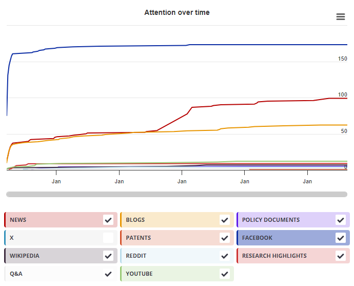
Timeline

Login to access the full chart related to this output.

If you don’t have an account, click here to discover Explorer

X Demographics

X Demographics

The data shown below were collected from the profiles of 10 X users who shared this research output. Click here to find out more about how the information was compiled.
As of 1 July 2024, you may notice a temporary increase in the numbers of X profiles with Unknown location. Click here to learn more.
Mendeley readers

Mendeley readers

The data shown below were compiled from readership statistics for 58 Mendeley readers of this research output. Click here to see the associated Mendeley record.

Geographical breakdown

Country Count As %
Netherlands 1 2%
Unknown 57 98%

Demographic breakdown

Readers by professional status Count As %
Student > Ph. D. Student 9 16%
Student > Master 9 16%
Student > Bachelor 8 14%
Student > Doctoral Student 4 7%
Researcher 4 7%
Other 10 17%
Unknown 14 24%
Readers by discipline Count As %
Neuroscience 19 33%
Agricultural and Biological Sciences 10 17%
Biochemistry, Genetics and Molecular Biology 5 9%
Medicine and Dentistry 3 5%
Nursing and Health Professions 1 2%
Other 4 7%
Unknown 16 28%
Attention Score in Context

Attention Score in Context

This research output has an Altmetric Attention Score of 5. This is our high-level measure of the quality and quantity of online attention that it has received. This Attention Score, as well as the ranking and number of research outputs shown below, was calculated when the research output was last mentioned on 06 July 2015.
All research outputs
#6,961,098
of 25,182,110 outputs
Outputs from Frontiers in Neuroanatomy
#397
of 1,250 outputs
Outputs of similar age
#73,844
of 269,274 outputs
Outputs of similar age from Frontiers in Neuroanatomy
#16
of 50 outputs
Altmetric has tracked 25,182,110 research outputs across all sources so far. This one has received more attention than most of these and is in the 72nd percentile.
So far Altmetric has tracked 1,250 research outputs from this source. They typically receive more attention than average, with a mean Attention Score of 9.0. This one has gotten more attention than average, scoring higher than 67% of its peers.
Older research outputs will score higher simply because they've had more time to accumulate mentions. To account for age we can compare this Altmetric Attention Score to the 269,274 tracked outputs that were published within six weeks on either side of this one in any source. This one has gotten more attention than average, scoring higher than 72% of its contemporaries.
We're also able to compare this research output to 50 others from the same source and published within six weeks on either side of this one. This one has gotten more attention than average, scoring higher than 68% of its contemporaries.