↓ Skip to main content

From Structure to Circuits: The Contribution of MEG Connectivity Studies to Functional Neurosurgery

Overview of attention for article published in Frontiers in Neuroanatomy, June 2016
Altmetric Badge

About this Attention Score

  • Average Attention Score compared to outputs of the same age
  • Average Attention Score compared to outputs of the same age and source

Mentioned by

twitter
4 X users

Citations

dimensions_citation
18 Dimensions

Readers on

mendeley
69 Mendeley
You are seeing a free-to-access but limited selection of the activity Altmetric has collected about this research output. Click here to find out more.
Title
From Structure to Circuits: The Contribution of MEG Connectivity Studies to Functional Neurosurgery
Published in
Frontiers in Neuroanatomy, June 2016
DOI 10.3389/fnana.2016.00067
Pubmed ID
Authors

Elizabeth W. Pang, O. C. Snead

Abstract

New advances in structural neuroimaging have revealed the intricate and extensive connections within the brain, data which have informed a number of ambitious projects such as the mapping of the human connectome. Elucidation of the structural connections of the brain, at both the macro and micro levels, promises new perspectives on brain structure and function that could translate into improved outcomes in functional neurosurgery. The understanding of neuronal structural connectivity afforded by these data now offers a vista on the brain, in both healthy and diseased states, that could not be seen with traditional neuroimaging. Concurrent with these developments in structural imaging, a complementary modality called magnetoencephalography (MEG) has been garnering great attention because it too holds promise for being able to shed light on the intricacies of functional brain connectivity. MEG is based upon the elemental principle of physics that an electrical current generates a magnetic field. Hence, MEG uses highly sensitive biomagnetometers to measure extracranial magnetic fields produced by intracellular neuronal currents. Put simply then, MEG is a measure of neurophysiological activity, which captures the magnetic fields generated by synchronized intraneuronal electrical activity. As such, MEG recordings offer exquisite resolution in the time and oscillatory domain and, as well, when co-registered with magnetic resonance imaging (MRI), offer excellent resolution in the spatial domain. Recent advances in MEG computational and graph theoretical methods have led to studies of connectivity in the time-frequency domain. As such, MEG can elucidate a neurophysiological-based functional circuitry that may enhance what is seen with MRI connectivity studies. In particular, MEG may offer additional insight not possible by MRI when used to study complex eloquent function, where the precise timing and coordination of brain areas is critical. This article will review the traditional use of MEG for functional neurosurgery, describe recent advances in MEG connectivity analyses, and consider the additional benefits that could be gained with the inclusion of MEG connectivity studies. Since MEG has been most widely applied to the study of epilepsy, we will frame this article within the context of epilepsy surgery and functional neurosurgery for epilepsy.
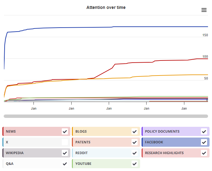
Timeline

Login to access the full chart related to this output.

If you don’t have an account, click here to discover Explorer

X Demographics

X Demographics

The data shown below were collected from the profiles of 4 X users who shared this research output. Click here to find out more about how the information was compiled.
As of 1 July 2024, you may notice a temporary increase in the numbers of X profiles with Unknown location. Click here to learn more.
Mendeley readers

Mendeley readers

The data shown below were compiled from readership statistics for 69 Mendeley readers of this research output. Click here to see the associated Mendeley record.

Geographical breakdown

Country Count As %
United Kingdom 1 1%
Unknown 68 99%

Demographic breakdown

Readers by professional status Count As %
Researcher 9 13%
Student > Master 9 13%
Student > Bachelor 9 13%
Student > Ph. D. Student 7 10%
Other 6 9%
Other 17 25%
Unknown 12 17%
Readers by discipline Count As %
Neuroscience 16 23%
Medicine and Dentistry 14 20%
Engineering 9 13%
Psychology 4 6%
Agricultural and Biological Sciences 3 4%
Other 6 9%
Unknown 17 25%
Attention Score in Context

Attention Score in Context

This research output has an Altmetric Attention Score of 2. This is our high-level measure of the quality and quantity of online attention that it has received. This Attention Score, as well as the ranking and number of research outputs shown below, was calculated when the research output was last mentioned on 30 June 2016.
All research outputs
#13,781,256
of 22,879,161 outputs
Outputs from Frontiers in Neuroanatomy
#603
of 1,162 outputs
Outputs of similar age
#192,552
of 353,105 outputs
Outputs of similar age from Frontiers in Neuroanatomy
#15
of 30 outputs
Altmetric has tracked 22,879,161 research outputs across all sources so far. This one is in the 38th percentile – i.e., 38% of other outputs scored the same or lower than it.
So far Altmetric has tracked 1,162 research outputs from this source. They typically receive more attention than average, with a mean Attention Score of 8.9. This one is in the 47th percentile – i.e., 47% of its peers scored the same or lower than it.
Older research outputs will score higher simply because they've had more time to accumulate mentions. To account for age we can compare this Altmetric Attention Score to the 353,105 tracked outputs that were published within six weeks on either side of this one in any source. This one is in the 44th percentile – i.e., 44% of its contemporaries scored the same or lower than it.
We're also able to compare this research output to 30 others from the same source and published within six weeks on either side of this one. This one has gotten more attention than average, scoring higher than 50% of its contemporaries.