↓ Skip to main content

The Isotropic Fractionator as a Tool for Quantitative Analysis in Central Nervous System Diseases

Overview of attention for article published in Frontiers in Cellular Neuroscience, August 2016
Altmetric Badge

Mentioned by

twitter
1 X user

Citations

dimensions_citation
13 Dimensions

Readers on

mendeley
30 Mendeley
You are seeing a free-to-access but limited selection of the activity Altmetric has collected about this research output. Click here to find out more.
Title
The Isotropic Fractionator as a Tool for Quantitative Analysis in Central Nervous System Diseases
Published in
Frontiers in Cellular Neuroscience, August 2016
DOI 10.3389/fncel.2016.00190
Pubmed ID
Authors

Ivan E. Repetto, Riccardo Monti, Marta Tropiano, Simone Tomasi, Alessia Arbini, Carlos-Humberto Andrade-Moraes, Roberto Lent, Alessandro Vercelli

Abstract

One major aim in quantitative and translational neuroscience is to achieve a precise and fast neuronal counting method to work on high throughput scale to obtain reliable results. Here, we tested the isotropic fractionator (IF) method for evaluating neuronal and non-neuronal cell loss in different models of central nervous system (CNS) pathologies. Sprague-Dawley rats underwent: (i) ischemic brain damage; (ii) intraperitoneal injection with kainic acid (KA) to induce epileptic seizures; and (iii) monolateral striatal injection with quinolinic acid (QA) mimicking human Huntington's disease. All specimens were processed for IF method and cell loss assessed. Hippocampus from KA-treated rats and striatum from QA-treated rats were carefully dissected using a dissection microscope and a rat brain matrix. Ischemic rat brains slices were first processed for TTC staining and then for IF. In the ischemic group the cell loss corresponded to the neuronal loss suggesting that hypoxia primarily affects neurons. Combining IF with TTC staining we could correlate the volume of lesion to the neuronal loss; by IF, we could assess that neuronal loss also occurs contralaterally to the ischemic side. In the epileptic group we observed a reduction of neuronal cells in treated rats, but also evaluated the changes in the number of non-neuronal cells in response to the hippocampal damage. In the QA model, there was a robust reduction of neuronal cells on ipsilateral striatum. This neuronal cell loss was not related to a drastic change in the total number of cells, being overcome by the increase in non-neuronal cells, thus suggesting that excitotoxic damage in the striatum strongly activates inflammation and glial proliferation. We concluded that the IF method could represent a simple and reliable quantitative technique to evaluate the effects of experimental lesions mimicking human diseases, and to consider the neuroprotective/anti-inflammatory effects of different treatments in the whole brain and also in discrete regions of interest, with the potential to investigate non-neuronal alterations. Moreover, IF could be used in addition or in substitution to classical stereological techniques or TTC staining used so far, since it is fast, precise and easily combined with complex molecular analysis.
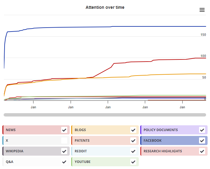
Timeline

Login to access the full chart related to this output.

If you don’t have an account, click here to discover Explorer

X Demographics

X Demographics

The data shown below were collected from the profile of 1 X user who shared this research output. Click here to find out more about how the information was compiled.
As of 1 July 2024, you may notice a temporary increase in the numbers of X profiles with Unknown location. Click here to learn more.
Mendeley readers

Mendeley readers

The data shown below were compiled from readership statistics for 30 Mendeley readers of this research output. Click here to see the associated Mendeley record.

Geographical breakdown

Country Count As %
Unknown 30 100%

Demographic breakdown

Readers by professional status Count As %
Student > Ph. D. Student 5 17%
Other 4 13%
Student > Doctoral Student 4 13%
Researcher 3 10%
Student > Master 3 10%
Other 9 30%
Unknown 2 7%
Readers by discipline Count As %
Neuroscience 11 37%
Agricultural and Biological Sciences 5 17%
Biochemistry, Genetics and Molecular Biology 3 10%
Medicine and Dentistry 2 7%
Psychology 1 3%
Other 5 17%
Unknown 3 10%
Attention Score in Context

Attention Score in Context

This research output has an Altmetric Attention Score of 1. This is our high-level measure of the quality and quantity of online attention that it has received. This Attention Score, as well as the ranking and number of research outputs shown below, was calculated when the research output was last mentioned on 05 August 2016.
All research outputs
#20,336,685
of 22,881,964 outputs
Outputs from Frontiers in Cellular Neuroscience
#3,586
of 4,256 outputs
Outputs of similar age
#321,923
of 366,897 outputs
Outputs of similar age from Frontiers in Cellular Neuroscience
#44
of 53 outputs
Altmetric has tracked 22,881,964 research outputs across all sources so far. This one is in the 1st percentile – i.e., 1% of other outputs scored the same or lower than it.
So far Altmetric has tracked 4,256 research outputs from this source. They typically receive a little more attention than average, with a mean Attention Score of 6.2. This one is in the 1st percentile – i.e., 1% of its peers scored the same or lower than it.
Older research outputs will score higher simply because they've had more time to accumulate mentions. To account for age we can compare this Altmetric Attention Score to the 366,897 tracked outputs that were published within six weeks on either side of this one in any source. This one is in the 1st percentile – i.e., 1% of its contemporaries scored the same or lower than it.
We're also able to compare this research output to 53 others from the same source and published within six weeks on either side of this one. This one is in the 1st percentile – i.e., 1% of its contemporaries scored the same or lower than it.