↓ Skip to main content

Brain Morphometry and the Neurobiology of Levodopa-Induced Dyskinesias: Current Knowledge and Future Potential for Translational Pre-Clinical Neuroimaging Studies

Overview of attention for article published in Frontiers in Neurology, June 2014
Altmetric Badge

About this Attention Score

  • Average Attention Score compared to outputs of the same age
  • Above-average Attention Score compared to outputs of the same age and source (64th percentile)

Mentioned by

twitter
3 X users

Citations

dimensions_citation
26 Dimensions

Readers on

mendeley
66 Mendeley
You are seeing a free-to-access but limited selection of the activity Altmetric has collected about this research output. Click here to find out more.
Title
Brain Morphometry and the Neurobiology of Levodopa-Induced Dyskinesias: Current Knowledge and Future Potential for Translational Pre-Clinical Neuroimaging Studies
Published in
Frontiers in Neurology, June 2014
DOI 10.3389/fneur.2014.00095
Pubmed ID
Authors

Clare J. Finlay, Susan Duty, Anthony C. Vernon

Abstract

Dopamine replacement therapy in the form of levodopa results in a significant proportion of patients with Parkinson's disease developing debilitating dyskinesia. This significantly complicates further treatment and negatively impacts patient quality of life. A greater understanding of the neurobiological mechanisms underlying levodopa-induced dyskinesia (LID) is therefore crucial to develop new treatments to prevent or mitigate LID. Such investigations in humans are largely confined to assessment of neurochemical and cerebrovascular blood flow changes using positron emission tomography and functional magnetic resonance imaging. However, recent evidence suggests that LID is associated with specific morphological changes in the frontal cortex and midbrain, detectable by structural MRI and voxel-based morphometry. Current human neuroimaging methods however lack sufficient resolution to reveal the biological mechanism driving these morphological changes at the cellular level. In contrast, there is a wealth of literature from well-established rodent models of LID documenting detailed post-mortem cellular and molecular measurements. The combination therefore of advanced neuroimaging methods and rodent LID models offers an exciting opportunity to bridge these currently disparate areas of research. To highlight this opportunity, in this mini-review, we provide an overview of the current clinical evidence for morphological changes in the brain associated with LID and identify potential cellular mechanisms as suggested from human and animal studies. We then suggest a framework for combining small animal MRI imaging with rodent models of LID, which may provide important mechanistic insights into the neurobiology of LID.
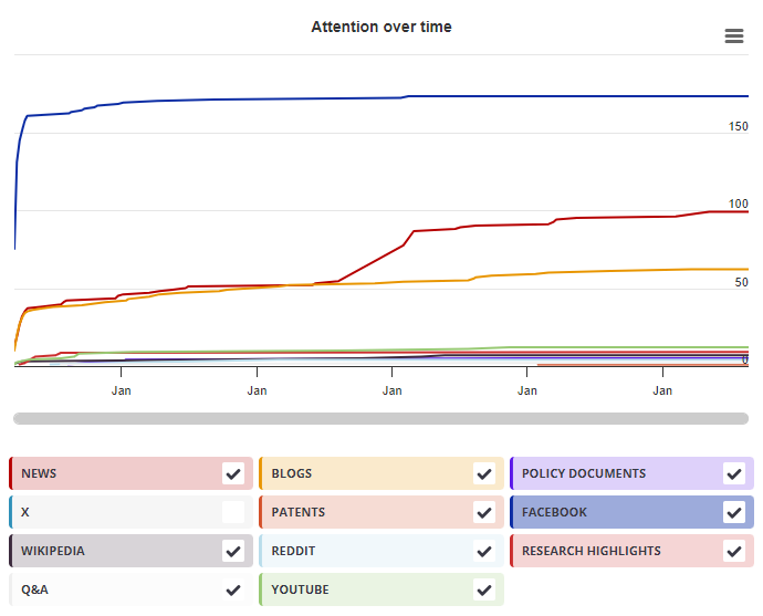
Timeline

Login to access the full chart related to this output.

If you don’t have an account, click here to discover Explorer

X Demographics

X Demographics

The data shown below were collected from the profiles of 3 X users who shared this research output. Click here to find out more about how the information was compiled.
As of 1 July 2024, you may notice a temporary increase in the numbers of X profiles with Unknown location. Click here to learn more.
Mendeley readers

Mendeley readers

The data shown below were compiled from readership statistics for 66 Mendeley readers of this research output. Click here to see the associated Mendeley record.

Geographical breakdown

Country Count As %
United States 1 2%
Unknown 65 98%

Demographic breakdown

Readers by professional status Count As %
Researcher 15 23%
Student > Ph. D. Student 13 20%
Student > Master 8 12%
Student > Bachelor 7 11%
Professor > Associate Professor 4 6%
Other 11 17%
Unknown 8 12%
Readers by discipline Count As %
Agricultural and Biological Sciences 16 24%
Medicine and Dentistry 12 18%
Neuroscience 11 17%
Biochemistry, Genetics and Molecular Biology 4 6%
Psychology 4 6%
Other 6 9%
Unknown 13 20%
Attention Score in Context

Attention Score in Context

This research output has an Altmetric Attention Score of 2. This is our high-level measure of the quality and quantity of online attention that it has received. This Attention Score, as well as the ranking and number of research outputs shown below, was calculated when the research output was last mentioned on 05 August 2014.
All research outputs
#14,781,727
of 22,757,090 outputs
Outputs from Frontiers in Neurology
#6,061
of 11,665 outputs
Outputs of similar age
#127,458
of 228,693 outputs
Outputs of similar age from Frontiers in Neurology
#21
of 68 outputs
Altmetric has tracked 22,757,090 research outputs across all sources so far. This one is in the 32nd percentile – i.e., 32% of other outputs scored the same or lower than it.
So far Altmetric has tracked 11,665 research outputs from this source. They typically receive a little more attention than average, with a mean Attention Score of 7.3. This one is in the 43rd percentile – i.e., 43% of its peers scored the same or lower than it.
Older research outputs will score higher simply because they've had more time to accumulate mentions. To account for age we can compare this Altmetric Attention Score to the 228,693 tracked outputs that were published within six weeks on either side of this one in any source. This one is in the 41st percentile – i.e., 41% of its contemporaries scored the same or lower than it.
We're also able to compare this research output to 68 others from the same source and published within six weeks on either side of this one. This one has gotten more attention than average, scoring higher than 64% of its contemporaries.