↓ Skip to main content

Outer Plexiform Layer Structures Are Not Altered Following AAV-Mediated Gene Transfer in Healthy Rat Retina

Overview of attention for article published in Frontiers in Neurology, February 2017
Altmetric Badge

Mentioned by

twitter
1 X user

Citations

dimensions_citation
2 Dimensions

Readers on

mendeley
24 Mendeley
You are seeing a free-to-access but limited selection of the activity Altmetric has collected about this research output. Click here to find out more.
Title
Outer Plexiform Layer Structures Are Not Altered Following AAV-Mediated Gene Transfer in Healthy Rat Retina
Published in
Frontiers in Neurology, February 2017
DOI 10.3389/fneur.2017.00059
Pubmed ID
Authors

Bert C. Giers, Daniela Klein, Alexandra Mendes-Madeira, Carolina Isiegas, Birgit Lorenz, Silke Haverkamp, Knut Stieger

Abstract

Ocular gene therapy approaches have been developed for a variety of different diseases. In particular, clinical gene therapy trials for RPE65 mutations, X-linked retinoschisis, and choroideremia have been conducted at different centers in recent years, showing that adeno-associated virus (AAV)-mediated gene therapy is safe, but limitations exist as to the therapeutic benefit and long-term duration of the treatment. The technique of vector delivery to retinal cells relies on subretinal injection of the vector solution, causing a transient retinal detachment. Although retinal detachments are known to cause remodeling of retinal neuronal structures as well as significant cell loss, the possible effects of this short-term therapeutic retinal detachment on retinal structure and circuitry have not yet been studied in detail. In this study, retinal morphology and apoptotic status were examined in healthy rat retinas following AAV-mediated gene transfer via subretinal injection with AAV2/5.CMV.d2GFP or sham injection with fluorescein. Outer plexiform layer (OPL) morphology was assessed by immunohistochemical labeling, laser scanning confocal microscopy, and electron microscopy. The number of synaptic contacts in the OPL was quantified after labeling with structural markers. To assess the apoptotic status, inflammatory and pro-apoptotic markers were tested and TUNEL assay for the detection of apoptotic nuclei was performed. Pre- and postsynaptic structures in the OPL, such as synaptic ribbons or horizontal and bipolar cell processes, did not differ in size or shape in injected versus non-injected areas and control retinas. Absolute numbers of synaptic ribbons were not altered. No signs of relevant gliosis were detected. TUNEL labeling of retinal cells did not vary between injected and non-injected areas, and apoptosis-inducing factor was not delocalized to the nucleus in transduced areas. The neuronal circuits in the OPL of healthy rat retinas undergoing AAV-mediated gene transfer were not altered by the temporary retinal detachment caused by subretinal injection, the presence of viral particles, or the expression of green fluorescent protein as a transgene. This observation likely requires further investigations in the dog model for RPE65 deficiency in order to determine the impact of RPE65 transgene expression on diseased retinas in animals and men.
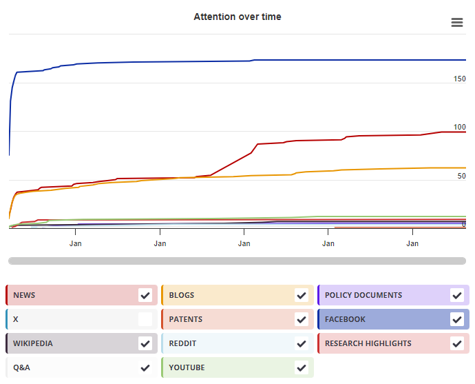
Timeline

Login to access the full chart related to this output.

If you don’t have an account, click here to discover Explorer

X Demographics

X Demographics

The data shown below were collected from the profile of 1 X user who shared this research output. Click here to find out more about how the information was compiled.
As of 1 July 2024, you may notice a temporary increase in the numbers of X profiles with Unknown location. Click here to learn more.
Mendeley readers

Mendeley readers

The data shown below were compiled from readership statistics for 24 Mendeley readers of this research output. Click here to see the associated Mendeley record.

Geographical breakdown

Country Count As %
Unknown 24 100%

Demographic breakdown

Readers by professional status Count As %
Researcher 9 38%
Student > Doctoral Student 1 4%
Student > Bachelor 1 4%
Other 1 4%
Student > Ph. D. Student 1 4%
Other 3 13%
Unknown 8 33%
Readers by discipline Count As %
Biochemistry, Genetics and Molecular Biology 4 17%
Neuroscience 4 17%
Medicine and Dentistry 3 13%
Agricultural and Biological Sciences 2 8%
Veterinary Science and Veterinary Medicine 1 4%
Other 1 4%
Unknown 9 38%
Attention Score in Context

Attention Score in Context

This research output has an Altmetric Attention Score of 1. This is our high-level measure of the quality and quantity of online attention that it has received. This Attention Score, as well as the ranking and number of research outputs shown below, was calculated when the research output was last mentioned on 23 February 2017.
All research outputs
#20,406,219
of 22,955,959 outputs
Outputs from Frontiers in Neurology
#8,864
of 11,842 outputs
Outputs of similar age
#271,173
of 311,210 outputs
Outputs of similar age from Frontiers in Neurology
#104
of 130 outputs
Altmetric has tracked 22,955,959 research outputs across all sources so far. This one is in the 1st percentile – i.e., 1% of other outputs scored the same or lower than it.
So far Altmetric has tracked 11,842 research outputs from this source. They typically receive a little more attention than average, with a mean Attention Score of 7.3. This one is in the 1st percentile – i.e., 1% of its peers scored the same or lower than it.
Older research outputs will score higher simply because they've had more time to accumulate mentions. To account for age we can compare this Altmetric Attention Score to the 311,210 tracked outputs that were published within six weeks on either side of this one in any source. This one is in the 1st percentile – i.e., 1% of its contemporaries scored the same or lower than it.
We're also able to compare this research output to 130 others from the same source and published within six weeks on either side of this one. This one is in the 1st percentile – i.e., 1% of its contemporaries scored the same or lower than it.