↓ Skip to main content

Arsenic Exposure Induces Unscheduled Mitotic S Phase Entry Coupled with Cell Death in Mouse Cortical Astrocytes

Overview of attention for article published in Frontiers in Neuroscience, June 2016
Altmetric Badge

About this Attention Score

  • Average Attention Score compared to outputs of the same age
  • Average Attention Score compared to outputs of the same age and source

Mentioned by

twitter
5 X users

Citations

dimensions_citation
10 Dimensions

Readers on

mendeley
23 Mendeley
You are seeing a free-to-access but limited selection of the activity Altmetric has collected about this research output. Click here to find out more.
Title
Arsenic Exposure Induces Unscheduled Mitotic S Phase Entry Coupled with Cell Death in Mouse Cortical Astrocytes
Published in
Frontiers in Neuroscience, June 2016
DOI 10.3389/fnins.2016.00297
Pubmed ID
Authors

Nang T. T. Htike, Fumihiko Maekawa, Haruka Soutome, Kazuhiro Sano, Sho Maejima, Kyaw H. Aung, Masaaki Tokuda, Shinji Tsukahara

Abstract

There is serious concern about arsenic in the natural environment, which exhibits neurotoxicity and increases the risk of neurodevelopmental disorders. Adverse effects of arsenic have been demonstrated in neurons, but it is not fully understood how arsenic affects other cell types in the brain. In the current study, we examined whether sodium arsenite (NaAsO2) affects the cell cycle, viability, and apoptosis of in vitro-cultured astrocytes isolated from the cerebral cortex of mice. Cultured astrocytes from transgenic mice expressing fluorescent ubiquitination-based cell cycle indicator (Fucci) were subjected to live imaging analysis to assess the effects of NaAsO2 (0, 1, 2, and 4 μM) on the cell cycle and number of cells. Fucci was designed to express monomeric Kusabira Orange2 (mKO2) fused with the ubiquitylation domain of hCdt1, a marker of G1 phase, and monomeric Azami Green (mAG) fused with the ubiquitylation domain of hGem, a marker of S, G2, and M phases. NaAsO2 concentration-dependently decreased the peak levels of the mAG/mKO2 emission ratio when the ratio had reached a peak in astrocytes without NaAsO2 exposure, which was due to attenuating the increase in the mAG-expressing cell number. In contrast, the mAG/mKO2 emission ratio and number of mAG-expressing cells were concentration-dependently increased by NaAsO2 before their peak levels, indicating unscheduled S phase entry. We further examined the fate of cells forced to enter S phase by NaAsO2. We found that most of these cells died up to the end of live imaging. In addition, quantification of the copy number of the glial fibrillary acidic protein gene expressed specifically in astrocytes revealed a concentration-dependent decrease caused by NaAsO2. However, NaAsO2 did not increase the amount of nucleosomes generated from DNA fragmentation and failed to alter the gene expression of molecules relevant to unscheduled S phase entry-coupled apoptosis (p21, p53, E2F1, E2F4, and Gm36566). These findings suggest that NaAsO2 adversely affects the cell cycle and viability of astrocytes by inducing unscheduled S phase entry coupled with cell death that may be caused by mechanisms other than apoptosis.
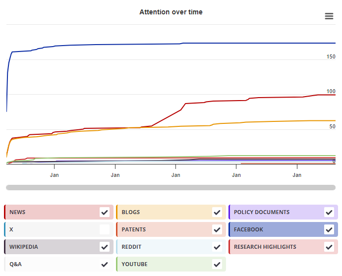
Timeline

Login to access the full chart related to this output.

If you don’t have an account, click here to discover Explorer

X Demographics

X Demographics

The data shown below were collected from the profiles of 5 X users who shared this research output. Click here to find out more about how the information was compiled.
As of 1 July 2024, you may notice a temporary increase in the numbers of X profiles with Unknown location. Click here to learn more.
Mendeley readers

Mendeley readers

The data shown below were compiled from readership statistics for 23 Mendeley readers of this research output. Click here to see the associated Mendeley record.

Geographical breakdown

Country Count As %
Unknown 23 100%

Demographic breakdown

Readers by professional status Count As %
Student > Ph. D. Student 5 22%
Student > Master 3 13%
Student > Doctoral Student 2 9%
Student > Bachelor 2 9%
Lecturer 1 4%
Other 4 17%
Unknown 6 26%
Readers by discipline Count As %
Agricultural and Biological Sciences 5 22%
Medicine and Dentistry 3 13%
Neuroscience 3 13%
Biochemistry, Genetics and Molecular Biology 2 9%
Unspecified 1 4%
Other 1 4%
Unknown 8 35%
Attention Score in Context

Attention Score in Context

This research output has an Altmetric Attention Score of 2. This is our high-level measure of the quality and quantity of online attention that it has received. This Attention Score, as well as the ranking and number of research outputs shown below, was calculated when the research output was last mentioned on 25 July 2016.
All research outputs
#15,739,529
of 25,373,627 outputs
Outputs from Frontiers in Neuroscience
#6,686
of 11,538 outputs
Outputs of similar age
#210,061
of 367,290 outputs
Outputs of similar age from Frontiers in Neuroscience
#96
of 156 outputs
Altmetric has tracked 25,373,627 research outputs across all sources so far. This one is in the 37th percentile – i.e., 37% of other outputs scored the same or lower than it.
So far Altmetric has tracked 11,538 research outputs from this source. They typically receive a lot more attention than average, with a mean Attention Score of 10.9. This one is in the 39th percentile – i.e., 39% of its peers scored the same or lower than it.
Older research outputs will score higher simply because they've had more time to accumulate mentions. To account for age we can compare this Altmetric Attention Score to the 367,290 tracked outputs that were published within six weeks on either side of this one in any source. This one is in the 41st percentile – i.e., 41% of its contemporaries scored the same or lower than it.
We're also able to compare this research output to 156 others from the same source and published within six weeks on either side of this one. This one is in the 33rd percentile – i.e., 33% of its contemporaries scored the same or lower than it.