↓ Skip to main content

Clinical Effects and Differences in Neural Function Connectivity Revealed by MRI in Subacute Hemispheric and Brainstem Infarction Patients With Dysphagia After Swallowing Therapy

Overview of attention for article published in Frontiers in Neuroscience, July 2018
Altmetric Badge

About this Attention Score

  • Average Attention Score compared to outputs of the same age
  • Average Attention Score compared to outputs of the same age and source

Mentioned by

twitter
4 X users

Readers on

mendeley
49 Mendeley
You are seeing a free-to-access but limited selection of the activity Altmetric has collected about this research output. Click here to find out more.
Title
Clinical Effects and Differences in Neural Function Connectivity Revealed by MRI in Subacute Hemispheric and Brainstem Infarction Patients With Dysphagia After Swallowing Therapy
Published in
Frontiers in Neuroscience, July 2018
DOI 10.3389/fnins.2018.00488
Pubmed ID
Authors

Yu-Chi Huang, Tun-Wei Hsu, Chau-Peng Leong, Han-Chin Hsieh, Wei-Che Lin

Abstract

Background: Early detection and intervention for post-stroke dysphagia could reduce the incidence of pulmonary complications and mortality. The aims of this study were to investigate the benefits of swallowing therapy in swallowing function and brain neuro-plasticity and to explore the relationship between swallowing function recovery and neuroplasticity after swallowing therapy in cerebral and brainstem stroke patients with dysphagia. Methods: We collected 17 subacute stroke patients with dysphagia (11 cerebral stroke patients with a median age of 76 years and 6 brainstem stroke patients with a median age of 70 years). Each patient received swallowing therapies during hospitalization. For each patient, functional oral intake scale (FOIS), functional dysphagia scale (FDS) and 8-point penetration-aspiration scale (PAS) in videofluoroscopy swallowing study (VFSS), and brain functional magnetic resonance imaging (fMRI) were evaluated before and after treatment. Results: FOIS (p = 0.003 in hemispheric group and p = 0.039 in brainstem group) and FDS (p = 0.006 in hemispheric group and p = 0.028 in brainstem group) were both significantly improved after treatment in hemispheric and brainstem stroke patients. In hemispheric stroke patients, changes in FOIS were related to changes of functional brain connectivity in the ventral default mode network (vDMN) of the precuneus in brain functional MRI (fMRI). In brainstem stroke patients, changes in FOIS were related to changes of functional brain connectivity in the left sensorimotor network (LSMN) of the left postcentral region characterized by brain fMRI. Conclusion: Both hemispheric and brainstem stroke patients with different swallowing difficulties showed improvements after swallowing training. For these two dysphagic stroke groups with corresponding etiologies, swallowing therapy could contribute to different functional neuroplasticity.
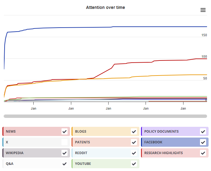
Timeline

Login to access the full chart related to this output.

If you don’t have an account, click here to discover Explorer

X Demographics

X Demographics

The data shown below were collected from the profiles of 4 X users who shared this research output. Click here to find out more about how the information was compiled.
As of 1 July 2024, you may notice a temporary increase in the numbers of X profiles with Unknown location. Click here to learn more.
Mendeley readers

Mendeley readers

The data shown below were compiled from readership statistics for 49 Mendeley readers of this research output. Click here to see the associated Mendeley record.

Geographical breakdown

Country Count As %
Unknown 49 100%

Demographic breakdown

Readers by professional status Count As %
Student > Master 7 14%
Researcher 6 12%
Student > Ph. D. Student 6 12%
Student > Bachelor 5 10%
Professor > Associate Professor 3 6%
Other 6 12%
Unknown 16 33%
Readers by discipline Count As %
Nursing and Health Professions 15 31%
Medicine and Dentistry 5 10%
Neuroscience 3 6%
Unspecified 2 4%
Pharmacology, Toxicology and Pharmaceutical Science 1 2%
Other 4 8%
Unknown 19 39%
Attention Score in Context

Attention Score in Context

This research output has an Altmetric Attention Score of 2. This is our high-level measure of the quality and quantity of online attention that it has received. This Attention Score, as well as the ranking and number of research outputs shown below, was calculated when the research output was last mentioned on 30 July 2018.
All research outputs
#15,175,718
of 25,385,509 outputs
Outputs from Frontiers in Neuroscience
#6,404
of 11,542 outputs
Outputs of similar age
#180,127
of 340,079 outputs
Outputs of similar age from Frontiers in Neuroscience
#148
of 233 outputs
Altmetric has tracked 25,385,509 research outputs across all sources so far. This one is in the 38th percentile – i.e., 38% of other outputs scored the same or lower than it.
So far Altmetric has tracked 11,542 research outputs from this source. They typically receive a lot more attention than average, with a mean Attention Score of 11.0. This one is in the 42nd percentile – i.e., 42% of its peers scored the same or lower than it.
Older research outputs will score higher simply because they've had more time to accumulate mentions. To account for age we can compare this Altmetric Attention Score to the 340,079 tracked outputs that were published within six weeks on either side of this one in any source. This one is in the 45th percentile – i.e., 45% of its contemporaries scored the same or lower than it.
We're also able to compare this research output to 233 others from the same source and published within six weeks on either side of this one. This one is in the 33rd percentile – i.e., 33% of its contemporaries scored the same or lower than it.