↓ Skip to main content

Callosal Influence on Visual Receptive Fields Has an Ocular, an Orientation-and Direction Bias

Overview of attention for article published in Frontiers in Systems Neuroscience, April 2018
Altmetric Badge

About this Attention Score

  • Average Attention Score compared to outputs of the same age

Mentioned by

twitter
3 X users

Citations

dimensions_citation
10 Dimensions

Readers on

mendeley
27 Mendeley
You are seeing a free-to-access but limited selection of the activity Altmetric has collected about this research output. Click here to find out more.
Title
Callosal Influence on Visual Receptive Fields Has an Ocular, an Orientation-and Direction Bias
Published in
Frontiers in Systems Neuroscience, April 2018
DOI 10.3389/fnsys.2018.00011
Pubmed ID
Authors

Sergio A. Conde-Ocazionez, Christiane Jungen, Thomas Wunderle, David Eriksson, Sergio Neuenschwander, Kerstin E. Schmidt

Abstract

One leading hypothesis on the nature of visual callosal connections (CC) is that they replicate features of intrahemispheric lateral connections. However, CC act also in the central part of the binocular visual field. In agreement, early experiments in cats indicated that they provide the ipsilateral eye part of binocular receptive fields (RFs) at the vertical midline (Berlucchi and Rizzolatti, 1968), and play a key role in stereoscopic function. But until today callosal inputs to receptive fields activated by one or both eyes were never compared simultaneously, because callosal function has been often studied by cutting or lesioning either corpus callosum or optic chiasm not allowing such a comparison. To investigate the functional contribution of CC in the intact cat visual system we recorded both monocular and binocular neuronal spiking responses and receptive fields in the 17/18 transition zone during reversible deactivation of the contralateral hemisphere. Unexpectedly from many of the previous reports, we observe no change in ocular dominance during CC deactivation. Throughout the transition zone, a majority of RFs shrink, but several also increase in size. RFs are significantly more affected for ipsi- as opposed to contralateral stimulation, but changes are also observed with binocular stimulation. Noteworthy, RF shrinkages are tiny and not correlated to the profound decreases of monocular and binocular firing rates. They depend more on orientation and direction preference than on eccentricity or ocular dominance of the receiving neuron's RF. Our findings confirm that in binocularly viewing mammals, binocular RFs near the midline are constructed via the direct geniculo-cortical pathway. They also support the idea that input from the two eyes complement each other through CC: Rather than linking parts of RFs separated by the vertical meridian, CC convey a modulatory influence, reflecting the feature selectivity of lateral circuits, with a strong cardinal bias.
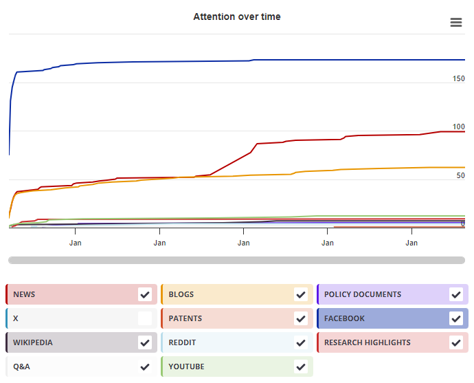
Timeline

Login to access the full chart related to this output.

If you don’t have an account, click here to discover Explorer

X Demographics

X Demographics

The data shown below were collected from the profiles of 3 X users who shared this research output. Click here to find out more about how the information was compiled.
As of 1 July 2024, you may notice a temporary increase in the numbers of X profiles with Unknown location. Click here to learn more.
Mendeley readers

Mendeley readers

The data shown below were compiled from readership statistics for 27 Mendeley readers of this research output. Click here to see the associated Mendeley record.

Geographical breakdown

Country Count As %
Unknown 27 100%

Demographic breakdown

Readers by professional status Count As %
Researcher 9 33%
Student > Bachelor 4 15%
Student > Ph. D. Student 3 11%
Student > Master 3 11%
Professor 2 7%
Other 2 7%
Unknown 4 15%
Readers by discipline Count As %
Neuroscience 15 56%
Agricultural and Biological Sciences 5 19%
Arts and Humanities 1 4%
Nursing and Health Professions 1 4%
Medicine and Dentistry 1 4%
Other 1 4%
Unknown 3 11%
Attention Score in Context

Attention Score in Context

This research output has an Altmetric Attention Score of 2. This is our high-level measure of the quality and quantity of online attention that it has received. This Attention Score, as well as the ranking and number of research outputs shown below, was calculated when the research output was last mentioned on 21 April 2018.
All research outputs
#14,970,944
of 23,028,364 outputs
Outputs from Frontiers in Systems Neuroscience
#891
of 1,345 outputs
Outputs of similar age
#179,693
of 296,837 outputs
Outputs of similar age from Frontiers in Systems Neuroscience
#13
of 19 outputs
Altmetric has tracked 23,028,364 research outputs across all sources so far. This one is in the 32nd percentile – i.e., 32% of other outputs scored the same or lower than it.
So far Altmetric has tracked 1,345 research outputs from this source. They typically receive a lot more attention than average, with a mean Attention Score of 10.7. This one is in the 28th percentile – i.e., 28% of its peers scored the same or lower than it.
Older research outputs will score higher simply because they've had more time to accumulate mentions. To account for age we can compare this Altmetric Attention Score to the 296,837 tracked outputs that were published within six weeks on either side of this one in any source. This one is in the 36th percentile – i.e., 36% of its contemporaries scored the same or lower than it.
We're also able to compare this research output to 19 others from the same source and published within six weeks on either side of this one. This one is in the 26th percentile – i.e., 26% of its contemporaries scored the same or lower than it.