↓ Skip to main content

Retinal Afferents Synapse with Relay Cells Targeting the Middle Temporal Area in the Pulvinar and Lateral Geniculate Nuclei

Overview of attention for article published in Frontiers in Neuroanatomy, January 2010
Altmetric Badge

About this Attention Score

  • Good Attention Score compared to outputs of the same age (75th percentile)
  • Good Attention Score compared to outputs of the same age and source (66th percentile)

Mentioned by

twitter
2 X users
wikipedia
4 Wikipedia pages

Citations

dimensions_citation
129 Dimensions

Readers on

mendeley
104 Mendeley
You are seeing a free-to-access but limited selection of the activity Altmetric has collected about this research output. Click here to find out more.
Title
Retinal Afferents Synapse with Relay Cells Targeting the Middle Temporal Area in the Pulvinar and Lateral Geniculate Nuclei
Published in
Frontiers in Neuroanatomy, January 2010
DOI 10.3389/neuro.05.008.2010
Pubmed ID
Authors

Claire E. Warner, Yona Goldshmit, James A. Bourne

Abstract

Considerable debate continues regarding thalamic inputs to the middle temporal area (MT) of the visual cortex that bypass the primary visual cortex (V1) and the role they might have in the residual visual capability following a lesion of V1. Two specific retinothalamic projections to area MT have been speculated to relay through the medial portion of the inferior pulvinar nucleus (PIm) and the koniocellular layers of the dorsal lateral geniculate nucleus (LGN). Although a number of studies have demonstrated retinal inputs to regions of the thalamus where relays to area MT have been observed, the relationship between the retinal terminals and area MT relay cells has not been established. Here we examined direct retino-recipient regions of the marmoset monkey (Callithrix jacchus) pulvinar nucleus and the LGN following binocular injections of anterograde tracer, as well as area MT relay cells in these nuclei by injection of retrograde tracer into area MT. Retinal afferents were shown to synapse with area MT relay cells as demonstrated by colocalization with the presynaptic vesicle membrane protein synaptophysin. We also established the presence of direct synapes of retinal afferents on area MT relay cells within the PIm, as well as the koniocellular K1 and K3 layers of the LGN, thereby corroborating the existence of two disynaptic pathways from the retina to area MT that bypass V1.
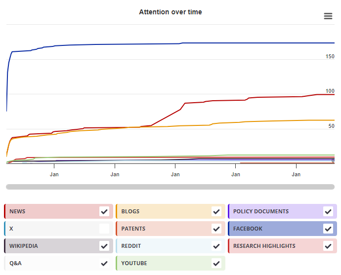
Timeline

Login to access the full chart related to this output.

If you don’t have an account, click here to discover Explorer

X Demographics

X Demographics

The data shown below were collected from the profiles of 2 X users who shared this research output. Click here to find out more about how the information was compiled.
As of 1 July 2024, you may notice a temporary increase in the numbers of X profiles with Unknown location. Click here to learn more.
Mendeley readers

Mendeley readers

The data shown below were compiled from readership statistics for 104 Mendeley readers of this research output. Click here to see the associated Mendeley record.

Geographical breakdown

Country Count As %
United Kingdom 2 2%
United States 2 2%
Canada 1 <1%
Japan 1 <1%
Spain 1 <1%
Unknown 97 93%

Demographic breakdown

Readers by professional status Count As %
Student > Ph. D. Student 29 28%
Researcher 20 19%
Student > Master 10 10%
Professor > Associate Professor 9 9%
Professor 6 6%
Other 14 13%
Unknown 16 15%
Readers by discipline Count As %
Neuroscience 31 30%
Agricultural and Biological Sciences 17 16%
Psychology 16 15%
Medicine and Dentistry 13 13%
Engineering 3 3%
Other 7 7%
Unknown 17 16%
Attention Score in Context

Attention Score in Context

This research output has an Altmetric Attention Score of 5. This is our high-level measure of the quality and quantity of online attention that it has received. This Attention Score, as well as the ranking and number of research outputs shown below, was calculated when the research output was last mentioned on 02 September 2022.
All research outputs
#6,280,735
of 23,230,825 outputs
Outputs from Frontiers in Neuroanatomy
#386
of 1,178 outputs
Outputs of similar age
#40,377
of 165,588 outputs
Outputs of similar age from Frontiers in Neuroanatomy
#6
of 18 outputs
Altmetric has tracked 23,230,825 research outputs across all sources so far. This one has received more attention than most of these and is in the 72nd percentile.
So far Altmetric has tracked 1,178 research outputs from this source. They typically receive more attention than average, with a mean Attention Score of 8.9. This one has gotten more attention than average, scoring higher than 66% of its peers.
Older research outputs will score higher simply because they've had more time to accumulate mentions. To account for age we can compare this Altmetric Attention Score to the 165,588 tracked outputs that were published within six weeks on either side of this one in any source. This one has done well, scoring higher than 75% of its contemporaries.
We're also able to compare this research output to 18 others from the same source and published within six weeks on either side of this one. This one has gotten more attention than average, scoring higher than 66% of its contemporaries.