↓ Skip to main content

Quantitative evaluation of fMRI retinotopic maps, from V1 to V4, for cognitive experiments

Overview of attention for article published in Frontiers in Human Neuroscience, May 2015
Altmetric Badge

About this Attention Score

  • In the top 25% of all research outputs scored by Altmetric
  • High Attention Score compared to outputs of the same age (87th percentile)
  • Good Attention Score compared to outputs of the same age and source (78th percentile)

Mentioned by

news
1 news outlet
twitter
6 X users

Citations

dimensions_citation
13 Dimensions

Readers on

mendeley
75 Mendeley
You are seeing a free-to-access but limited selection of the activity Altmetric has collected about this research output. Click here to find out more.
Title
Quantitative evaluation of fMRI retinotopic maps, from V1 to V4, for cognitive experiments
Published in
Frontiers in Human Neuroscience, May 2015
DOI 10.3389/fnhum.2015.00277
Pubmed ID
Authors

Cécile Bordier, Jean-Michel Hupé, Michel Dojat

Abstract

FMRI retinotopic mapping is a non-invasive technique for the delineation of low-level visual areas in individual subjects. It generally relies upon the analysis of functional responses to periodic visual stimuli that encode eccentricity or polar angle in the visual field. This technique is used in vision research when the precise assignation of brain activation to retinotopic areas is an issue. It involves processing steps computed with different algorithms and embedded in various software suites. Manual intervention may be needed for some steps. Although the diversity of the available processing suites and manual interventions may potentially introduce some differences in the final delineation of visual areas, no documented comparison between maps obtained with different procedures has been reported in the literature. To explore the effect of the processing steps on the quality of the maps obtained, we used two tools, BALC, which relies on a fully automated procedure, and BrainVoyager, where areas are delineated "by hand" on the brain surface. To focus on the mapping procedures specifically, we used the same SPM pipeline for pretreatment and the same tissue segmentation tool. We document the consistency and differences of the fMRI retinotopic maps obtained from "routine retinotopy" experiments on 10 subjects. The maps obtained by skilled users are never fully identical. However, the agreement between the maps, around 80% for low-level areas, is probably sufficient for most applications. Our results also indicate that assigning cognitive activations, following a specific experiment (here, color perception), to individual retinotopic maps is not free of errors. We provide measurements of this error, that may help for the cautious interpretation of cognitive activation projection onto fMRI retinotopic maps. On average, the magnitude of the error is about 20%, with much larger differences in a few subjects. More variability may even be expected with less trained users or using different acquisition parameters and preprocessing chains.
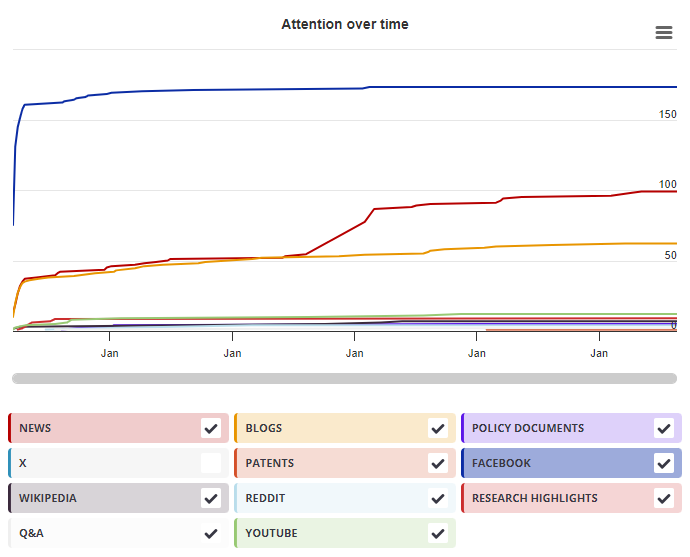
Timeline

Login to access the full chart related to this output.

If you don’t have an account, click here to discover Explorer

X Demographics

X Demographics

The data shown below were collected from the profiles of 6 X users who shared this research output. Click here to find out more about how the information was compiled.
As of 1 July 2024, you may notice a temporary increase in the numbers of X profiles with Unknown location. Click here to learn more.
Mendeley readers

Mendeley readers

The data shown below were compiled from readership statistics for 75 Mendeley readers of this research output. Click here to see the associated Mendeley record.

Geographical breakdown

Country Count As %
United Kingdom 1 1%
Netherlands 1 1%
Unknown 73 97%

Demographic breakdown

Readers by professional status Count As %
Researcher 18 24%
Student > Ph. D. Student 15 20%
Student > Master 7 9%
Student > Bachelor 6 8%
Student > Doctoral Student 6 8%
Other 9 12%
Unknown 14 19%
Readers by discipline Count As %
Neuroscience 21 28%
Psychology 12 16%
Medicine and Dentistry 7 9%
Agricultural and Biological Sciences 5 7%
Engineering 4 5%
Other 8 11%
Unknown 18 24%
Attention Score in Context

Attention Score in Context

This research output has an Altmetric Attention Score of 13. This is our high-level measure of the quality and quantity of online attention that it has received. This Attention Score, as well as the ranking and number of research outputs shown below, was calculated when the research output was last mentioned on 24 April 2016.
All research outputs
#2,911,719
of 26,160,558 outputs
Outputs from Frontiers in Human Neuroscience
#1,340
of 7,798 outputs
Outputs of similar age
#35,368
of 281,782 outputs
Outputs of similar age from Frontiers in Human Neuroscience
#41
of 188 outputs
Altmetric has tracked 26,160,558 research outputs across all sources so far. Compared to these this one has done well and is in the 88th percentile: it's in the top 25% of all research outputs ever tracked by Altmetric.
So far Altmetric has tracked 7,798 research outputs from this source. They typically receive a lot more attention than average, with a mean Attention Score of 15.1. This one has done well, scoring higher than 82% of its peers.
Older research outputs will score higher simply because they've had more time to accumulate mentions. To account for age we can compare this Altmetric Attention Score to the 281,782 tracked outputs that were published within six weeks on either side of this one in any source. This one has done well, scoring higher than 87% of its contemporaries.
We're also able to compare this research output to 188 others from the same source and published within six weeks on either side of this one. This one has done well, scoring higher than 78% of its contemporaries.