↓ Skip to main content

Cortical iron regulation and inflammatory response in Alzheimer's disease and APPSWE/PS1ΔE9 mice: a histological perspective

Overview of attention for article published in Frontiers in Neuroscience, July 2015
Altmetric Badge

About this Attention Score

  • In the top 25% of all research outputs scored by Altmetric
  • High Attention Score compared to outputs of the same age (84th percentile)
  • High Attention Score compared to outputs of the same age and source (83rd percentile)

Mentioned by

news
1 news outlet
twitter
2 X users

Readers on

mendeley
86 Mendeley
You are seeing a free-to-access but limited selection of the activity Altmetric has collected about this research output. Click here to find out more.
Title
Cortical iron regulation and inflammatory response in Alzheimer's disease and APPSWE/PS1ΔE9 mice: a histological perspective
Published in
Frontiers in Neuroscience, July 2015
DOI 10.3389/fnins.2015.00255
Pubmed ID
Authors

Mark D. Meadowcroft, James R. Connor, Qing X. Yang

Abstract

Disruption of iron homeostasis and increased glial response are known to occur in brains afflicted by Alzheimer's disease (AD). While the APP/PS1 transgenic mouse model recapitulates the hallmark amyloid-beta plaque pathology of AD, it does so in a different neuronal mileu than humans. Understanding the iron characteristics and glial response of the APP/PS1 model is important when testing new treatment procedures and translating these results. Brain tissue from AD patients, APP/PS1 mice, and controls were stained for iron, H- and L-ferritin, microglia, astrocytes, Aβ40∕42, and degenerating neurons. The histological data demonstrate differences in ferritin, iron distribution, gliosis, and Aβ plaque composition between APP/PS1 and AD tissue. Specifically, an association between focal iron deposition and Aβ plaques is found ubiquitously throughout the AD tissue and is not observed in the APP/PS1 mouse model. Ferritin, microglia, and astrocyte staining show differential response patterns to amyloid plaques in AD and the APP/PS1 tissue. Aβ 40 and 42 antibody and thioflavin staining demonstrate morphological differences in plaque composition. The histological data support the hypothesis that iron distribution, iron management, and glial response histologically differ between the APP/PS1 and AD brain. Acknowledging the caveat that there are distinct plaque, iron, and glial contrasts between the AD brain and the APP/PS1 mouse is crucial when utilizing this model.
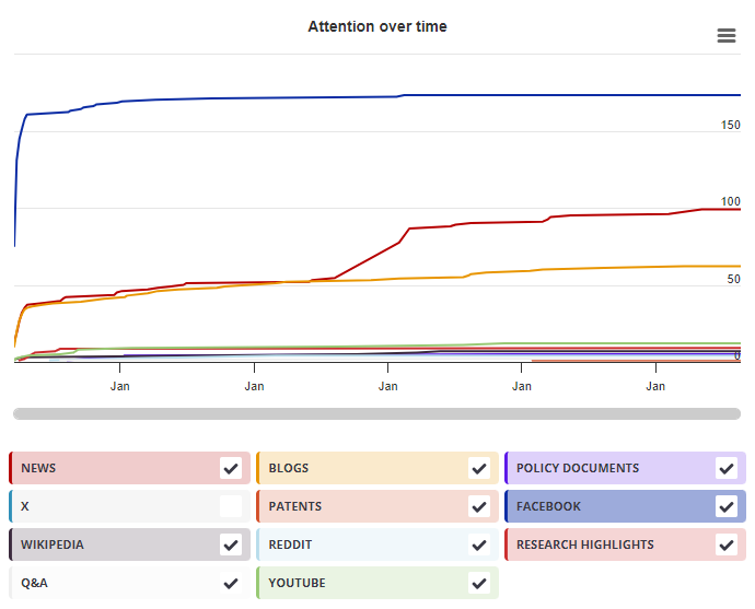
Timeline

Login to access the full chart related to this output.

If you don’t have an account, click here to discover Explorer

X Demographics

X Demographics

The data shown below were collected from the profiles of 2 X users who shared this research output. Click here to find out more about how the information was compiled.
As of 1 July 2024, you may notice a temporary increase in the numbers of X profiles with Unknown location. Click here to learn more.
Mendeley readers

Mendeley readers

The data shown below were compiled from readership statistics for 86 Mendeley readers of this research output. Click here to see the associated Mendeley record.

Geographical breakdown

Country Count As %
Germany 2 2%
Italy 1 1%
Unknown 83 97%

Demographic breakdown

Readers by professional status Count As %
Student > Ph. D. Student 20 23%
Researcher 12 14%
Student > Master 11 13%
Student > Bachelor 10 12%
Student > Doctoral Student 6 7%
Other 7 8%
Unknown 20 23%
Readers by discipline Count As %
Agricultural and Biological Sciences 19 22%
Neuroscience 17 20%
Biochemistry, Genetics and Molecular Biology 8 9%
Medicine and Dentistry 4 5%
Immunology and Microbiology 2 2%
Other 14 16%
Unknown 22 26%
Attention Score in Context

Attention Score in Context

This research output has an Altmetric Attention Score of 11. This is our high-level measure of the quality and quantity of online attention that it has received. This Attention Score, as well as the ranking and number of research outputs shown below, was calculated when the research output was last mentioned on 14 August 2015.
All research outputs
#3,394,510
of 25,374,917 outputs
Outputs from Frontiers in Neuroscience
#2,675
of 11,541 outputs
Outputs of similar age
#41,729
of 275,174 outputs
Outputs of similar age from Frontiers in Neuroscience
#17
of 107 outputs
Altmetric has tracked 25,374,917 research outputs across all sources so far. Compared to these this one has done well and is in the 86th percentile: it's in the top 25% of all research outputs ever tracked by Altmetric.
So far Altmetric has tracked 11,541 research outputs from this source. They typically receive a lot more attention than average, with a mean Attention Score of 10.9. This one has done well, scoring higher than 76% of its peers.
Older research outputs will score higher simply because they've had more time to accumulate mentions. To account for age we can compare this Altmetric Attention Score to the 275,174 tracked outputs that were published within six weeks on either side of this one in any source. This one has done well, scoring higher than 84% of its contemporaries.
We're also able to compare this research output to 107 others from the same source and published within six weeks on either side of this one. This one has done well, scoring higher than 83% of its contemporaries.